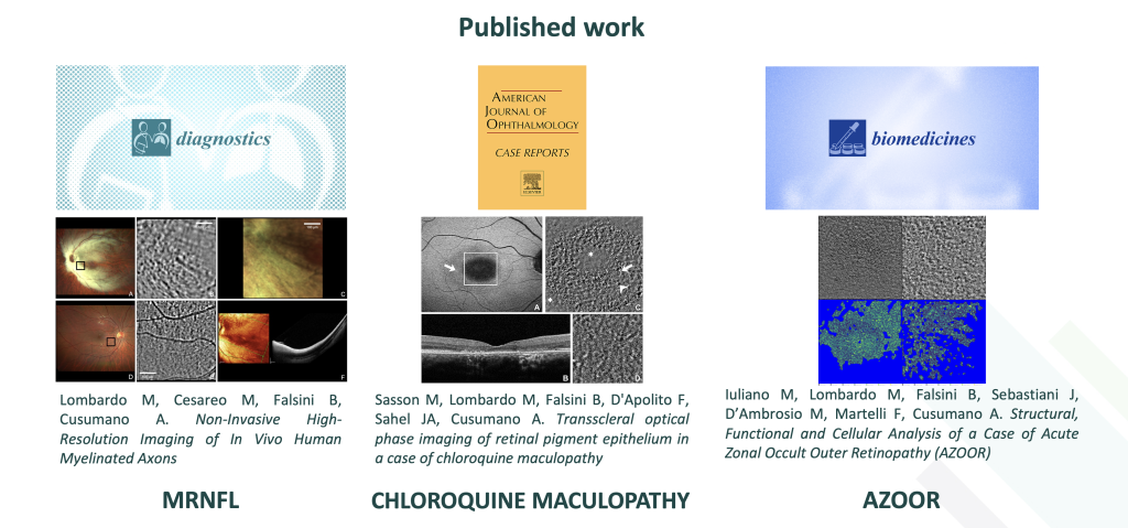

Over the past decades, continuous advances in retinal imaging techniques have enabled increasingly detailed visualization of retinal layers, allowing ophthalmologists to assess retinal health and identify structural alterations, tissue-level lesions, and damage. Yet many retinal diseases arise at the cellular level, long before classical clinical signs become evident, at a stage when irreversible damage may already have occurred and therapeutic interventions may be limited, less effective or even impossible.
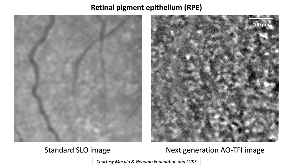
Our research leverages next-generation adaptive optics (AO) imaging to visualize the living retina with unprecedented resolution and contrast. This technology enables non-invasive, repeatable in vivo observation of critical retinal cells, such as photoreceptors and the retinal pigment epithelium (RPE) – a tissue essential for photoreceptor health and the first to be affected in many retinal diseases.
This is not merely improved imaging; it represents a step toward establishing cell-level biomarkers that can guide patient selection and timing optimization for emerging treatments through quantitative measures of retinal structure, with a level of precision that goes beyond the capabilities of conventional imaging. By moving from structural snapshots to measurable cellular metrics, we aim to redefine how retinal disease is detected, monitored, and treated.
Table of Contents
Project Overview
Next-generation adaptive optics combines advanced wavefront correction with innovative illumination strategies to overcome key limitations of conventional retinal imaging. One major breakthrough is transscleral illumination, which enhances signal and contrast for structures that are difficult to visualize through standard transpupillary approaches. This innovation allows visualization of layers that were previously inaccessible, providing a more complete understanding of retinal microstructure.
Using these techniques, we can capture cellular-level images over clinically relevant fields of view, with rapid acquisition times suitable for real-world clinical application. This capability makes AO imaging not only a research tool but also a platform ready for translational use in patient care.
Table of Contents
Why This Matters
The landscape of retinal therapy is evolving rapidly. Gene therapy, stem cell approaches, optogenetics, complement inhibitors for geographic atrophy, optopharmacology, and other emerging treatments are moving from experimental stages to clinical reality.
These innovations raise a critical question: how can we detect therapeutic need and effects early and accurately? Next-generation AO provides a solution by offering unprecedented insight into cellular-level retinal changes.
AO imaging allows clinicians and researchers to:
- Detect early cellular changes, often before photoreceptor damage occurs, non-invasively,
- Monitor therapy effects over time, non-invasively,
- Provide reproducible, quantitative endpoints suitable for clinical trials and regulatory evaluation.
The Scientific Breakthrough: Transscleral Adaptive Optics Imaging
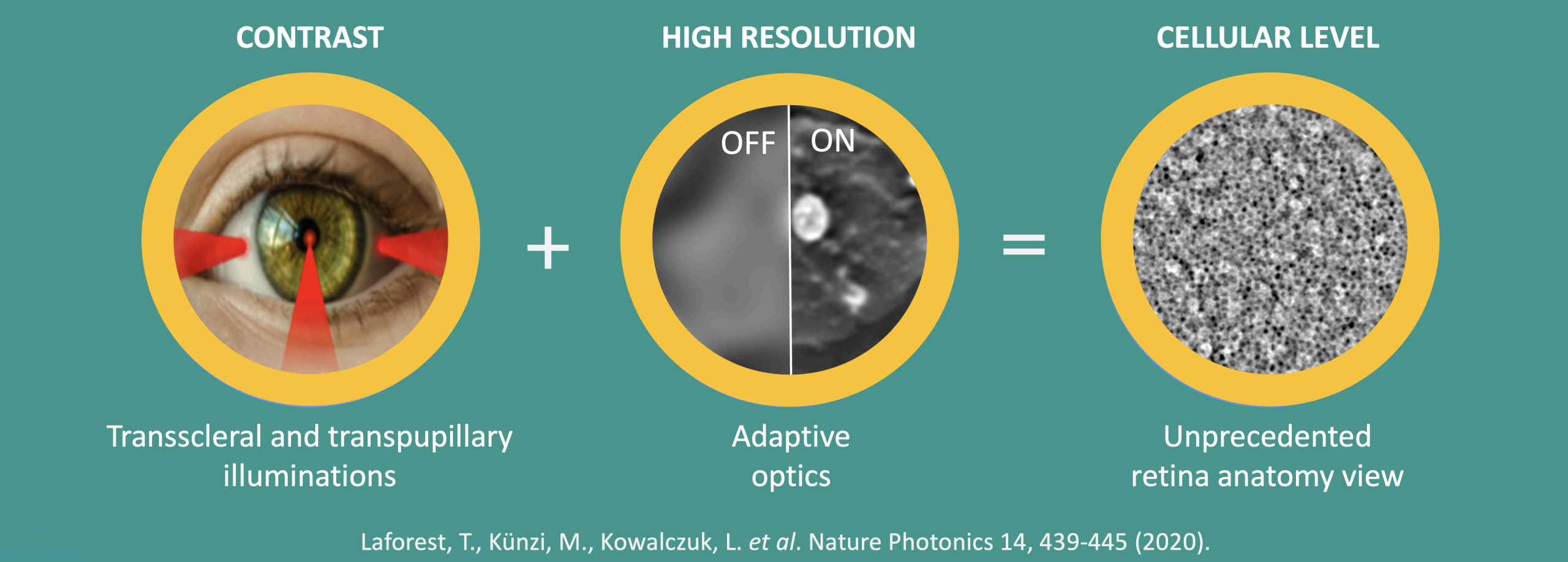
Conventional retinal imaging illuminates the eye through the pupil, which can limit visualization of certain cellular structures. Transscleral illumination offers an alternative pathway, improving light penetration and enhancing backscattered signals from deeper layers such as the RPE.
This approach, combined with adaptive optics, allows in vivo imaging of multiple retinal cell types with remarkable clarity. In healthy eyes, studies have demonstrated strong repeatability of cellular metrics – including cell density, area, and morphology – and identified correlations with age and axial length. These findings pave the way for normative databases and disease-specific deviations, which are critical for accurate diagnosis and monitoring.
From Images to Biomarkers
Our work focuses on transforming cellular images into quantitative metrics that can be tracked longitudinally and correlated with patient characteristics and disease progression.
Examples of cellular metrics include:
- Cell density and area (e.g., age-related changes in RPE cells),
- Morphology descriptors such as circularity, elongation, solidity, and border-distance dispersion,
- Nerve fiber layer structure and optic nerve head morphology, enabling early detection of optic neuropathies.
By identifying early structural changes at the cellular level, AO imaging can detect disease before irreversible photoreceptor or optic nerve damage occurs, enabling timely and potentially life-changing interventions.
Early Disease Applications
- Age-related macular degeneration (AMD) – early and late RPE alterations,
- Inherited retinal diseases (IRDs) and macular dystrophies,
- Central serous chorioretinopathy (CSCR) and other RPE/photoreceptor disorders,
- Retinal vascular disease and microcirculation abnormalities,
- Early nerve fiber layer changes relevant to glaucoma and other optic neuropathies.
Our long-term goal is to establish robust cellular biomarkers that support earlier diagnosis, enable precise monitoring of disease progression, and provide sensitive endpoints for evaluating therapeutic efficacy.
Clinical Relevance
For high-resolution retinal imaging to impact patient care, three elements are essential:
- Selecting the right patients,
- Timing interventions appropriately,
- Measuring structural outcomes with sensitive and repeatable endpoints.
Expected Impact
By advancing this research, we aim to accelerate the transition from macro-imaging to cell-level ophthalmology, enabling:
- Earlier and more precise detection of retinal cell alterations,
- Objective monitoring of disease progression and treatment response,
- Improved endpoints for clinical trials of novel therapies,
- A deeper understanding of the relationship between retinal microstructure and visual function.